Beyond diagnosis: is there a role for radiomics in prostate cancer management?
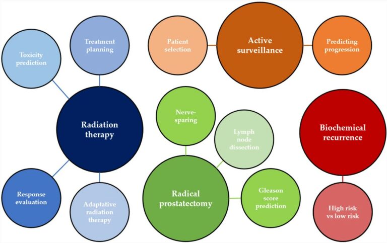
At present, therapeutic and prognostic recommendations for prostate cancer (PCa) predominantly hinge on risk-stratification tools that are built upon clinical parameters. Recent evidence indicates that incorporating imaging can enhance the precision of prognostic models based on clinical factors. However, challenges like subjective interpretation, variability in image analysis, and the absence of reliable quantitative measures need to be overcome to fully