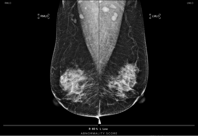
Implementing AI in breast imaging: challenges to turn the gadget into gain
In healthcare, the implementation of artificial intelligence (AI) is rapidly gaining momentum with breast imaging being no exception, or rather a poster child case. Numerous clinical indications intuitively lend themselves to AI enhancement. While the adoption of broad AI in clinical breast imaging practice has been more or less a silent revolution – it is already widely used for invite