image processing

Welcome to the blog on Artificial Intelligence of
the European Society of Radiology

This blog aims at bringing educational and critical perspectives on AI to readers. It should help imaging professionals to learn and keep up to date with the technologies being developed in this rapidly evolving field.

Most used hashtags:

Latest posts

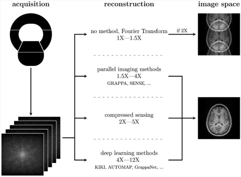
Complexities of deep learning-based undersampled MR image reconstruction

Recent advances in AI have led to deep learning-based MR undersampled image reconstruction methods showing more speed-ups compared to traditional algorithms. MR undersampling is an excellent way to reduce scan time but can negatively impact image quality. Our literature review aims to inform a broader audience about this complex topic. This highly multidisciplinary science requires the informed input of many

Read More →

AI for prostate MRI: open datasets, available applications, and grand challenges

This narrative review provides an overview of the current state-of-the-art artificial intelligence (AI) applications for prostate MRI by focusing on open datasets, commercially and publically available AI systems, and challenges. The authors state that large amounts of research are still required in order to successfully utilize AI in the whole prostate pathway. Due to the rapidly growing field, continuous up-to-date

Read More →

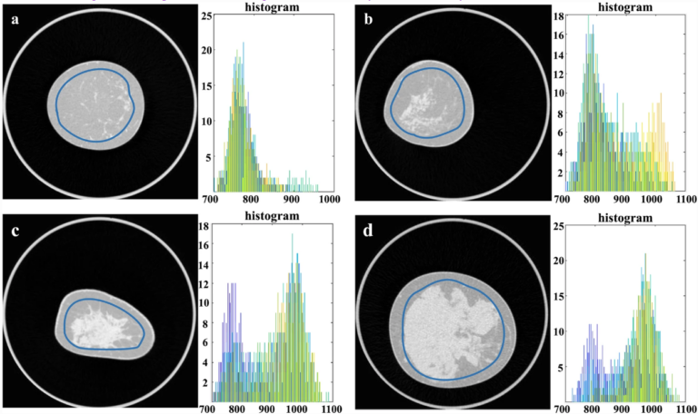
The potential of texture analysis for breast density classification

Breast cancer continues to be the most commonly diagnosed cancer among women with over 2 million new cases per year worldwide. One important independent risk factor for developing breast cancer is breast density (BD). Epidemiological studies show that women with dense tissue may have an increased risk of developing breast cancer by 2-6 times when compared to women with less

Read More →

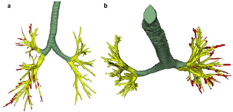
Creating a training set for AI from initial segmentations of airways

An important challenge in the use of artificial intelligence (AI) for medical image segmentation tasks is the lack of high-quality, scan protocol-specific datasets. AI performs best on narrow tasks with homogenous specifications. Thus, pre-trained models may be inadequate for use in centre-specific studies if the scan protocols do not match. For the airway segmentation task in the Imaging in Lifelines

Read More →

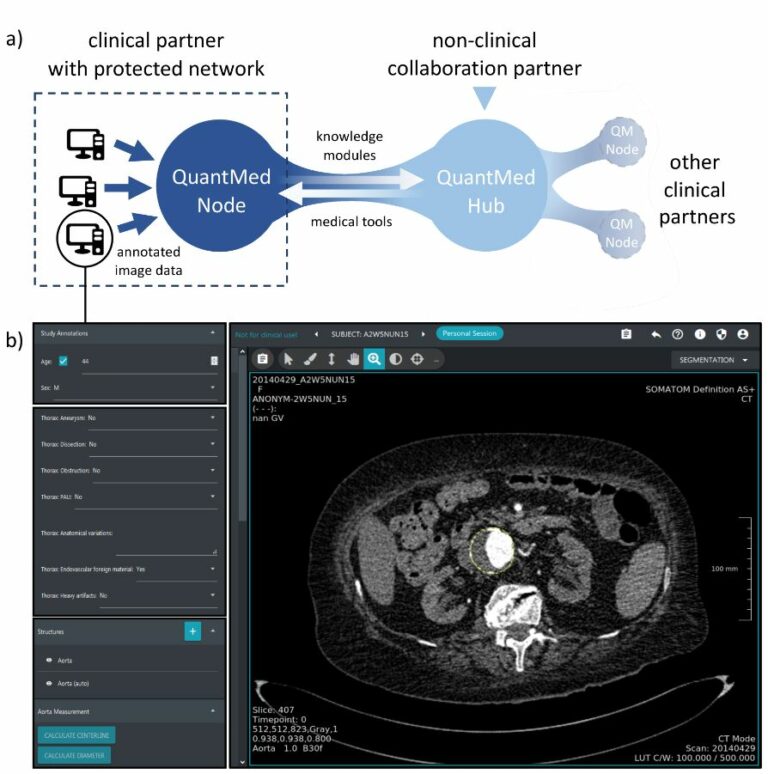
Automated segmentation and quantification of the healthy and diseased aorta in CT angiographies using a dedicated deep learning approach

Performed between 2015 and 2018, the purpose of this study was to develop and validate a deep learning-based algorithm for the segmentation and quantification of the physiological and diseased aorta in computed tomography (CT) angiographies. The authors were able to determine that automated segmentation of the aorta on CTA data using a deep learning algorithm is feasible and accurate, relating

Read More →

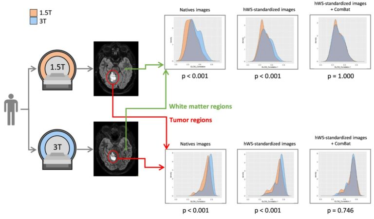
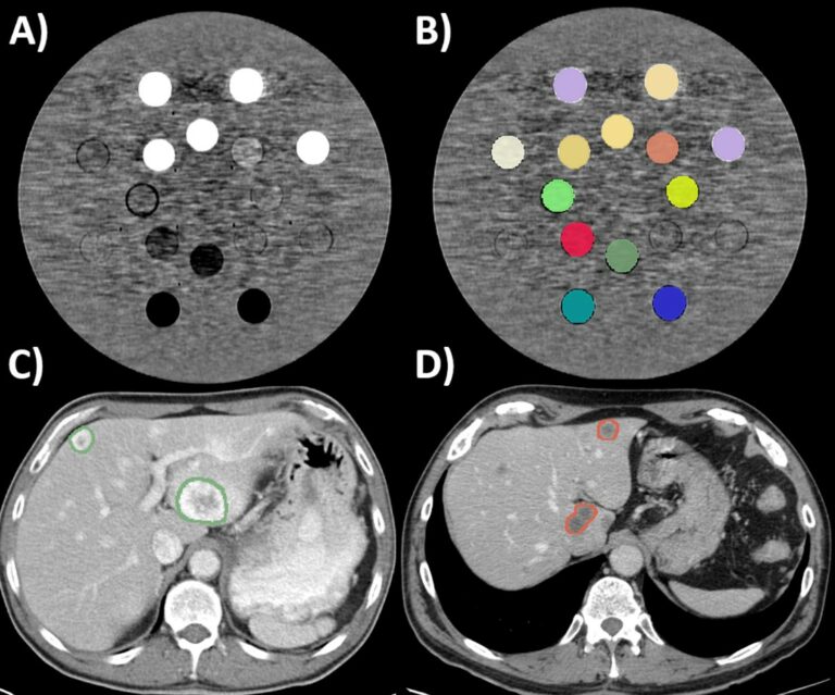
How can we combat multicenter variability in MR radiomics? Validation of a correction procedure

In this study, the authors extended the ComBat approach to provide a harmonization procedure that is applicable to any radiomic feature. They achieve this by combining image standardization with ComBat realignment, thus demonstrating that this could efficiently remove the scanner/protocol effect while preserving the individual variations in phantom, brain, and prostate MR scans. Key points Radiomic feature values obtained using

Read More →

Minimizing acquisition-related radiomics variability by image resampling and batch effect correction to allow for large-scale data analysis

Medical imaging encodes information of underlying tissues and can provide a comprehensive view of the entire body repeatedly throughout the course of disease. Therefore, medical imaging has been the foundation of disease detection and follow-up in clinical practice for decades. However, to date, observer-dependent evaluation of medical imaging has been a constraint for developing imaging biomarkers towards precision medicine. The

Read More →

Complexities of deep learning-based undersampled MR image reconstruction

Recent advances in AI have led to deep learning-based MR undersampled image reconstruction methods showing more speed-ups compared to traditional algorithms. MR undersampling is an excellent way to reduce scan time but can negatively impact image quality. Our literature review aims to inform a broader audience about this complex topic. This highly multidisciplinary science requires the informed input of many

Read More →

AI for prostate MRI: open datasets, available applications, and grand challenges

This narrative review provides an overview of the current state-of-the-art artificial intelligence (AI) applications for prostate MRI by focusing on open datasets, commercially and publically available AI systems, and challenges. The authors state that large amounts of research are still required in order to successfully utilize AI in the whole prostate pathway. Due to the rapidly growing field, continuous up-to-date

Read More →

The potential of texture analysis for breast density classification

Breast cancer continues to be the most commonly diagnosed cancer among women with over 2 million new cases per year worldwide. One important independent risk factor for developing breast cancer is breast density (BD). Epidemiological studies show that women with dense tissue may have an increased risk of developing breast cancer by 2-6 times when compared to women with less

Read More →

Creating a training set for AI from initial segmentations of airways

An important challenge in the use of artificial intelligence (AI) for medical image segmentation tasks is the lack of high-quality, scan protocol-specific datasets. AI performs best on narrow tasks with homogenous specifications. Thus, pre-trained models may be inadequate for use in centre-specific studies if the scan protocols do not match. For the airway segmentation task in the Imaging in Lifelines

Read More →

Automated segmentation and quantification of the healthy and diseased aorta in CT angiographies using a dedicated deep learning approach

Performed between 2015 and 2018, the purpose of this study was to develop and validate a deep learning-based algorithm for the segmentation and quantification of the physiological and diseased aorta in computed tomography (CT) angiographies. The authors were able to determine that automated segmentation of the aorta on CTA data using a deep learning algorithm is feasible and accurate, relating

Read More →

How can we combat multicenter variability in MR radiomics? Validation of a correction procedure

In this study, the authors extended the ComBat approach to provide a harmonization procedure that is applicable to any radiomic feature. They achieve this by combining image standardization with ComBat realignment, thus demonstrating that this could efficiently remove the scanner/protocol effect while preserving the individual variations in phantom, brain, and prostate MR scans. Key points Radiomic feature values obtained using

Read More →

Minimizing acquisition-related radiomics variability by image resampling and batch effect correction to allow for large-scale data analysis

Medical imaging encodes information of underlying tissues and can provide a comprehensive view of the entire body repeatedly throughout the course of disease. Therefore, medical imaging has been the foundation of disease detection and follow-up in clinical practice for decades. However, to date, observer-dependent evaluation of medical imaging has been a constraint for developing imaging biomarkers towards precision medicine. The

Read More →

Become A Member Today!

You will have access to a wide range of benefits that can help you advance your career and stay up-to-date with the latest developments in the field of radiology. These benefits include access to educational resources, networking opportunities with other professionals in the field, opportunities to participate in research projects and clinical trials, and access to the latest technologies and techniques. 

Check out our different membership options.

If you don’t find a fitting membership send us an email here.

Membership

for radiologists, radiology residents, professionals of allied sciences (including radiographers/radiological technologists, nuclear medicine physicians, medical physicists, and data scientists) & professionals of allied sciences in training residing within the boundaries of Europe

  • Reduced registration fees for ECR 1
  • Reduced fees for the European School of Radiology (ESOR) 2
  • Exclusive option to participate in the European Diploma. 3
  • Free electronic access to the journal European Radiology 4
  • Content e-mails for all ESR journals
  • Updates on offers & events through our newsletters
  • Exclusive access to the ESR feed in Juisci

€ 11 /year

Yes! That is less than €1 per month.

Free membership

for radiologists, radiology residents or professionals of allied sciences engaged in practice, teaching or research residing outside Europe as well as individual qualified professionals with an interest in radiology and medical imaging who do not fulfil individual or all requirements for any other ESR membership category & former full members who have retired from all clinical practice
  • Reduced registration fees for ECR 1
  • Free electronic access to the journal European Radiology
  • Content e-mails for all 3 ESR journals 4
  • Updates on offers & events through our newsletters
  • Exclusive access to the ESR feed in Juisci

€ 0

The best things in life are free.

ESR Friends

For students, company representatives or hospital managers etc.

  • Content e-mails for all 3 ESR journals 4
  • Updates on offers & events through our newsletters

€ 0

Friendship doesn’t cost a thing.

The membership type best fitting for you will be selected automatically during the application process.

Footnotes:

01

Reduced registration fees for ECR 2024:
Provided that ESR 2023 membership is activated and approved by August 31, 2023.

Reduced registration fees for ECR 2025:
Provided that ESR 2024 membership is activated and approved by August 31, 2024.

02
Not all activities included
03
Examination based on the ESR European Training Curriculum (radiologists or radiology residents).
04
European Radiology, Insights into Imaging, European Radiology Experimental.