CT

Welcome to the blog on Artificial Intelligence of
the European Society of Radiology

This blog aims at bringing educational and critical perspectives on AI to readers. It should help imaging professionals to learn and keep up to date with the technologies being developed in this rapidly evolving field.

Most used hashtags:

Latest posts

Deep learning assesses additional radiation dose in overscanning

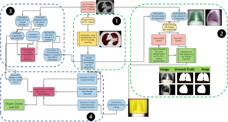
Following the COVID-19 pandemic, the number of chest CT examinations has dramatically increased, which will undeniably impact public medical exposure. Overscanning, i.e., scanning unnecessary regions in the axial field-of-view, causes noticeable excessive radiation dose to patients undergoing chest CT examinations. The manual procedure of selecting the scan range based on anterior-posterior or lateral localizers is prone to human error in

Read More →

New algorithm hopes to fight COVID-19 in international effort

The COVID-19 pandemic has seen doctors, healthcare institutions, and companies come together in an international effort to drive innovative solutions in the fight against this disease. AI-powered technology is one path that is being utilized to manage the heavy burden placed on clinicians as their workload increases. Dr. Dorin Comaniciu, who serves as Senior Vice President for Artificial Intelligence and

Read More →

Why radiomics research does not translate to clinical practice: evaluation of literature using RQS and TRIPOD

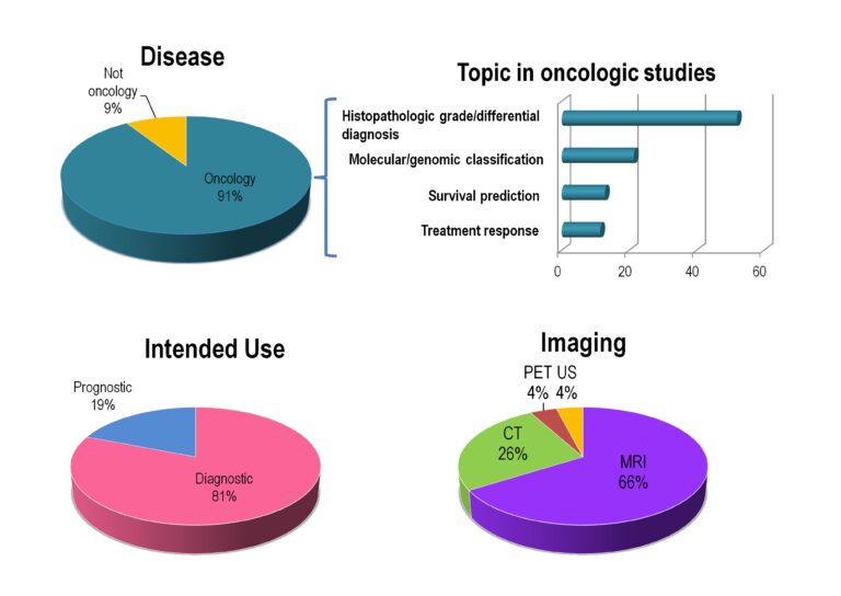
Over the last few years, the number of studies published using quantitative imaging biomarkers to classify or predict pathologies has steadily increased. As of today, a quick PubMed search for radiomics, imaging biomarkers or radiogenomics reveals well over 4,000 articles. However, somewhat surprisingly, given this amount of published research, outside of academic literature there is no widespread clinical application of

Read More →

Radiation dose in pregnancy

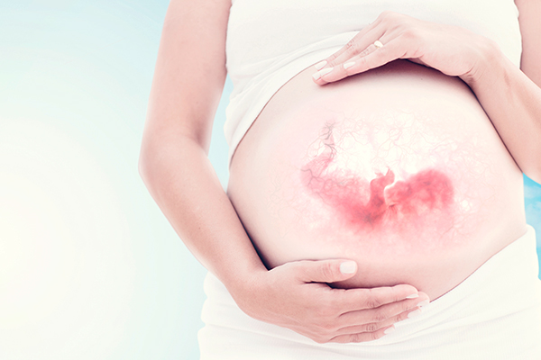
Due to the high radiosensitivity of the fetus and embryo, diagnostic imaging procedures for pregnant patients raise health concerns. Therefore, the authors of this work set out to develop a methodology for automated construction of patient-specific computational phantoms based on actual patient CT images with the aim of achieving accurate estimate of conceptus dose. Key points The conceptus dose during

Read More →

Can liver fibrosis be staged by deep learning techniques?

This pilot study aims to investigate whether liver fibrosis can be staged by deep learning techniques based on CT images. It included CT examinations of patients who underwent dynamic contrast-enhanced CT for evaluations of the liver and for whom histopathological information regarding liver fibrosis stage was available. Some additional images for training data were generated by rotating or parallel shifting

Read More →

AI for CT Image Reconstruction – A Great Opportunity

It seems like everybody in the radiology field is now working with Artificial Intelligence (AI) in one way or another. Especially here in the Silicon Valley, the number of ventures that develop AI algorithms is increasing by the day. What are these newly developed algorithms focusing on? Most of them try to solve a specific problem, and they do that

Read More →

Become A Member Today!

You will have access to a wide range of benefits that can help you advance your career and stay up-to-date with the latest developments in the field of radiology. These benefits include access to educational resources, networking opportunities with other professionals in the field, opportunities to participate in research projects and clinical trials, and access to the latest technologies and techniques. 

Check out our different membership options.

If you don’t find a fitting membership send us an email here.

Membership

for radiologists, radiology residents, professionals of allied sciences (including radiographers/radiological technologists, nuclear medicine physicians, medical physicists, and data scientists) & professionals of allied sciences in training residing within the boundaries of Europe

  • Reduced registration fees for ECR 1
  • Reduced fees for the European School of Radiology (ESOR) 2
  • Exclusive option to participate in the European Diploma. 3
  • Free electronic access to the journal European Radiology 4
  • Content e-mails for all ESR journals
  • Updates on offers & events through our newsletters
  • Exclusive access to the ESR feed in Juisci

€ 11 /year

Yes! That is less than €1 per month.

Free membership

for radiologists, radiology residents or professionals of allied sciences engaged in practice, teaching or research residing outside Europe as well as individual qualified professionals with an interest in radiology and medical imaging who do not fulfil individual or all requirements for any other ESR membership category & former full members who have retired from all clinical practice
  • Reduced registration fees for ECR 1
  • Free electronic access to the journal European Radiology
  • Content e-mails for all 3 ESR journals 4
  • Updates on offers & events through our newsletters
  • Exclusive access to the ESR feed in Juisci

€ 0

The best things in life are free.

ESR Friends

For students, company representatives or hospital managers etc.

  • Content e-mails for all 3 ESR journals 4
  • Updates on offers & events through our newsletters

€ 0

Friendship doesn’t cost a thing.

The membership type best fitting for you will be selected automatically during the application process.

Footnotes:

01

Reduced registration fees for ECR 2024:
Provided that ESR 2023 membership is activated and approved by August 31, 2023.

Reduced registration fees for ECR 2025:
Provided that ESR 2024 membership is activated and approved by August 31, 2024.

02
Not all activities included
03
Examination based on the ESR European Training Curriculum (radiologists or radiology residents).
04
European Radiology, Insights into Imaging, European Radiology Experimental.