Evaluating a deep learning software for lung parenchyma characterization in COVID-19 pneumonia
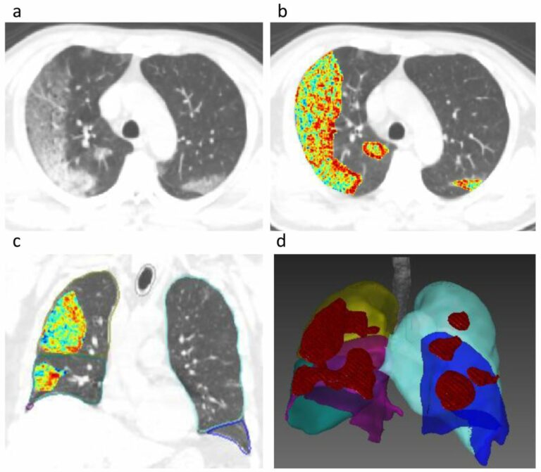
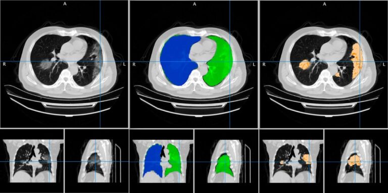
The aim of this study was to evaluate the performance of the LungQuant system, which is a deep learning-based software for quantitative analysis of chest CT. LungQuant was evaluated by comparing its results with independent visual evaluations by a group of clinical experts. The results indicated that an automatic quantification tool may be beneficial and contribute to an improved clinical