Fully automated convolutional neural network-based affine algorithm improves liver registration and lesion co-localization on hepatobiliary phase T1-weighted MR images
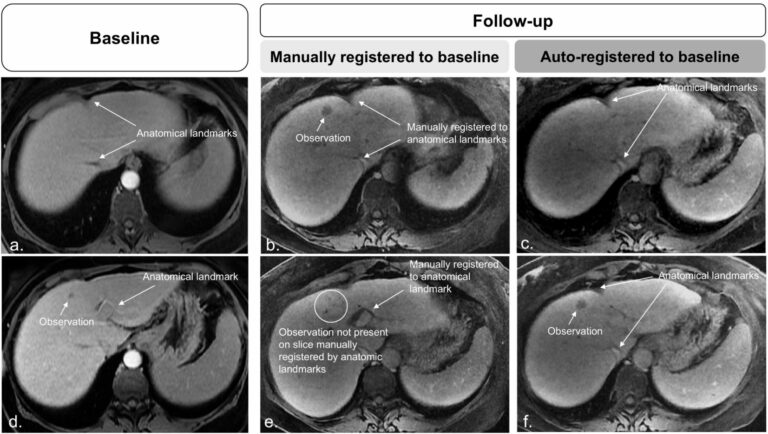
The authors of this study aimed to assess the performance of a convolutional neural network (CNN) algorithm to register cross-sectional liver imaging series and its performance to manual image registration. The study included three hundred fourteen patients who underwent gadoxetate disodium-enhanced magnetic resonance imaging (MRI) and were retrospectively selected. Key points Image registration across series can improve lesion co-localization and