algorithms

Welcome to the blog on Artificial Intelligence of
the European Society of Radiology

This blog aims at bringing educational and critical perspectives on AI to readers. It should help imaging professionals to learn and keep up to date with the technologies being developed in this rapidly evolving field.

Most used hashtags:

Latest posts

Radiologists’ performance improved by algorithmic transparency and interpretability measures

This study’s aim was to evaluate the perception of various types of artificial intelligence-based assistance and the interaction of radiologists with the algorithm’s predictions and certainty measures. As was consistent with previous research, the authors determined that human performance was superior to both groups when combined with AI. They also found an increase in trust in the algorithm’s performance when

Read More →

Machine learning–based radiomics classifies parotid tumors using morphological MRI

This comparative study aimed to evaluate the effectiveness of machine learning models based on morphological MRI radiomics in the classification of parotid tumors. The authors developed three-step machine learning models with extreme gradient boosting (XGBoost), support vector machine (SVM), and decision tree (DT) algorithms in order to classify the parotid neoplasms into four subtypes. The study was able to demonstrate

Read More →

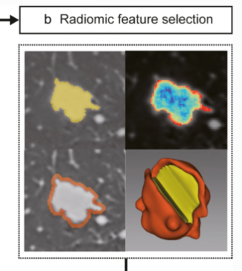
Radiomic nomogram predicts pathology invasiveness

The purpose of this retrospective diagnostic study was to develop and validate a preoperative CT-based nomogram combined with radiomic and clinical-radiological signatures to distinguish preinvasive lesions from pulmonary invasive lesions. The authors were able to demonstrate that a nomogram constructed by identified clinical-radiological signatures and combined radiomic signatures has the potential to precisely predict pathology invasiveness. Key points The radiomic

Read More →

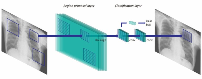
Optimal matrix size of chest radiographs for computer-aided detection on lung nodule or mass with deep learning

In this study, the authors retrospectively collected 2,088 abnormal and 352 normal chest radiographs from two institutions in order to investigate the optimal input matrix size for deep learning-based computer-aided detection (CAD) of nodules and masses on chest radiographs. This resulted in the matrix size 896 as having the highest performance for various sizes of abnormalities using different convolutional neural

Read More →

Data needs a human perspective

Artificial intelligence (AI) and machine learning have long since become part of our daily lives – as well as part of political discussions and programs. In healthcare, however, we should be especially careful with issues of bias, accountability, and privacy. Read the latest LinkedIn article from Siemens Healthineers CEO, Bernd Montag.

Read More →

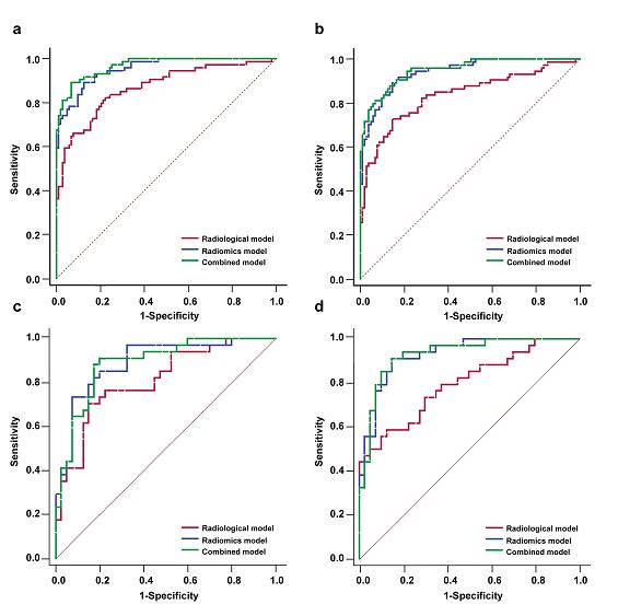
Noncontrast computer tomography–based radiomics model for predicting intracerebral hemorrhage expansion: preliminary findings and comparison with conventional radiological model

The authors developed a radiomics model for predicting hematoma expansion in patients with intracerebral haemorrhage (ICH) and compared its predictive performance with a conventional radiological feature-based model. Through retrospective analysis and noncontrast computed tomography (NCCT) assessment, it was found that an NCCT-based radiomics model showed better performance in the prediction of early hematoma expansion in ICH patients. Key points Radiomics

Read More →

Radiologists’ performance improved by algorithmic transparency and interpretability measures

This study’s aim was to evaluate the perception of various types of artificial intelligence-based assistance and the interaction of radiologists with the algorithm’s predictions and certainty measures. As was consistent with previous research, the authors determined that human performance was superior to both groups when combined with AI. They also found an increase in trust in the algorithm’s performance when

Read More →

Machine learning–based radiomics classifies parotid tumors using morphological MRI

This comparative study aimed to evaluate the effectiveness of machine learning models based on morphological MRI radiomics in the classification of parotid tumors. The authors developed three-step machine learning models with extreme gradient boosting (XGBoost), support vector machine (SVM), and decision tree (DT) algorithms in order to classify the parotid neoplasms into four subtypes. The study was able to demonstrate

Read More →

Radiomic nomogram predicts pathology invasiveness

The purpose of this retrospective diagnostic study was to develop and validate a preoperative CT-based nomogram combined with radiomic and clinical-radiological signatures to distinguish preinvasive lesions from pulmonary invasive lesions. The authors were able to demonstrate that a nomogram constructed by identified clinical-radiological signatures and combined radiomic signatures has the potential to precisely predict pathology invasiveness. Key points The radiomic

Read More →

Optimal matrix size of chest radiographs for computer-aided detection on lung nodule or mass with deep learning

In this study, the authors retrospectively collected 2,088 abnormal and 352 normal chest radiographs from two institutions in order to investigate the optimal input matrix size for deep learning-based computer-aided detection (CAD) of nodules and masses on chest radiographs. This resulted in the matrix size 896 as having the highest performance for various sizes of abnormalities using different convolutional neural

Read More →

Data needs a human perspective

Artificial intelligence (AI) and machine learning have long since become part of our daily lives – as well as part of political discussions and programs. In healthcare, however, we should be especially careful with issues of bias, accountability, and privacy. Read the latest LinkedIn article from Siemens Healthineers CEO, Bernd Montag.

Read More →

Noncontrast computer tomography–based radiomics model for predicting intracerebral hemorrhage expansion: preliminary findings and comparison with conventional radiological model

The authors developed a radiomics model for predicting hematoma expansion in patients with intracerebral haemorrhage (ICH) and compared its predictive performance with a conventional radiological feature-based model. Through retrospective analysis and noncontrast computed tomography (NCCT) assessment, it was found that an NCCT-based radiomics model showed better performance in the prediction of early hematoma expansion in ICH patients. Key points Radiomics

Read More →

Become A Member Today!

You will have access to a wide range of benefits that can help you advance your career and stay up-to-date with the latest developments in the field of radiology. These benefits include access to educational resources, networking opportunities with other professionals in the field, opportunities to participate in research projects and clinical trials, and access to the latest technologies and techniques. 

Check out our different membership options.

If you don’t find a fitting membership send us an email here.

Membership

for radiologists, radiology residents, professionals of allied sciences (including radiographers/radiological technologists, nuclear medicine physicians, medical physicists, and data scientists) & professionals of allied sciences in training residing within the boundaries of Europe

  • Reduced registration fees for ECR 1
  • Reduced fees for the European School of Radiology (ESOR) 2
  • Exclusive option to participate in the European Diploma. 3
  • Free electronic access to the journal European Radiology 4
  • Content e-mails for all ESR journals
  • Updates on offers & events through our newsletters
  • Exclusive access to the ESR feed in Juisci

€ 11 /year

Yes! That is less than €1 per month.

Free membership

for radiologists, radiology residents or professionals of allied sciences engaged in practice, teaching or research residing outside Europe as well as individual qualified professionals with an interest in radiology and medical imaging who do not fulfil individual or all requirements for any other ESR membership category & former full members who have retired from all clinical practice
  • Reduced registration fees for ECR 1
  • Free electronic access to the journal European Radiology
  • Content e-mails for all 3 ESR journals 4
  • Updates on offers & events through our newsletters
  • Exclusive access to the ESR feed in Juisci

€ 0

The best things in life are free.

ESR Friends

For students, company representatives or hospital managers etc.

  • Content e-mails for all 3 ESR journals 4
  • Updates on offers & events through our newsletters

€ 0

Friendship doesn’t cost a thing.

The membership type best fitting for you will be selected automatically during the application process.

Footnotes:

01

Reduced registration fees for ECR 2024:
Provided that ESR 2023 membership is activated and approved by August 31, 2023.

Reduced registration fees for ECR 2025:
Provided that ESR 2024 membership is activated and approved by August 31, 2024.

02
Not all activities included
03
Examination based on the ESR European Training Curriculum (radiologists or radiology residents).
04
European Radiology, Insights into Imaging, European Radiology Experimental.