“This was just awesome!” “Fantastic!” “Thank you for this great session!” These were only a few of the feedback messages in the chat at the end of the first episode of Reasons to do AI with Friends, entitled “The one where we classify breast cancer with MRI“.
Prof. Thomas Helbich, from the Medical University of Vienna, presented clinical evidence from his research group that contrast-enhanced MRI detects even small breast lesions, independent of the breast density. This modality has the ability to detect invasive, as well as non-invasive, cancers and has the highest sensitivity (>89%) in comparison to mammography or tomosynthesis and ultrasound (~40%). Helbich presented studies from his research group, and, in addition, from other international scientists of MRI screenings on women carrying a breast cancer gene and patients with an increased lifetime risk. Abbreviated multiparametric MRI, in particular, can obviate unnecessary breast biopsies, and is a fast, economical, highly beneficial and reliable technique.
The audience was able to follow closely via the chat and asked questions throughout the broadcasts, which the hosts answered live.
Prof. Helbich liaised with Prof. Georg Langs at the Medical University of Vienna in order to develop a reliable cancer screening machine learning (ML) tool that automatically detects, classifies and segments breast lesions. Langs explained the crucial requirements for such tools and how they can register, segment and classify even tiny lesions. The importance of the diversity of the patient set is underlined, which is crucial for the algorithm to learn how to discern between various lesion types. He pointed out that the structure of clinical data, as well as structured histology reports, are required to train the algorithm.
Helbich and Langs explained the ways in which radiologists and ML experts need to cooperate to create reliable algorithms. According to their findings, MRI is the better-suited modality, as it provides more information on the specifics of the lesion, more sequences and more parameters, which help to reduce the number of false positives. Data annotation and validation were explained in detail and how important the sample size and diversity of the data (e.g. different populations, centres and machines) are with regards to accuracy. The machine learns from the annotated imaging data and ML experts have to work in coordination with the radiologists to analyse and better understand the structure of the detected lesions. Moreover, validation of the data in the lab was covered in detail. A reduction of false positives and the number of biopsies should always be the aim when working with such ML tools.
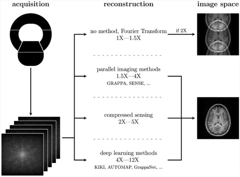
A highlight of the debut episode was Theo’s Technical Top Tips, a feature that will continue over the coming episodes, in which he presented the basic requirements when starting an ML project. Theodore Barfoot is an MRI physicist at the Royal Marsden London and a hobby beatboxer, showing off his skills with a rhythmical introduction to the clip that amazed the audience.
The chat was alive with activity throughout the broadcast, resulting in the hosts spending some extra time at the end of the webinar to make sure that all questions were answered.
Langs underlined that substituting breast imagers with AI devices would mean halting the state of the art where it is currently located, reassuring the viewers that he is convinced radiologists will always be required to advance present modalities.
The first episode of “AI Use Cases, reasons to do AI with friends” proved once again that live webinars can not only be informative but also entertaining. This season will run live every Wednesday until February 19 and present use cases for various issues. The next episode, “The one where we tackle fibrosis”, will be hosted by Helmut Prosch and Georg Langs from the Medical University of Vienna and will be broadcasted on January 22 at 19:30 CET. Registration is free of charge.
All episodes will be available for purchase on connect.myesr.org.
Read more on Episode 2, “The one where we tackle fibrosis” here.
Read more on Episode 3, “The one with the sixpack” here.
Read more on Episode 4, “The one that might be renal cancer” here.
Read more on Episode 5, “The one where we find the connection” here.
Read more on Episode 6, “The one with whole body MRI” here.
Want to deepen your AI knowledge? Check out the reading recommendations from our experts here.