


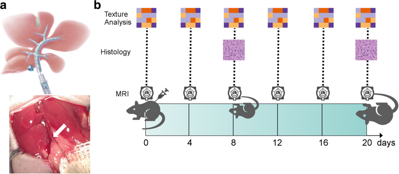
This study aimed to investigate whether any texture features show a correlation with intrahepatic tumor growth before the metastasis is visible to the human eye. For the purposes of the study, eight mice were injected intraportally with syngeneic MC-38 colon cancer cells and two mice were injected with phosphate-buffered saline (sham controls). Magnetic resonance imaging (MRI) and texture analysis were performed, and the features were examined with a linear regression model/Pearson correlation test and hierarchical cluster analysis. Tumors were visible on MRI after 20 days and 18 features from histogram and the gray-level-matrices exhibited statistically significant correlations before day 20 in the experiment group, but not in the control animals. The study concluded that texture features may quantitatively detect liver metastases before they become visually detectable by the radiologist.
Key points:
- Texture features change systematically in livers with (micro)metastases
- Three clusters of features independently correlated with tumor growth
- Texture features may quantitatively detect hepatic micrometastases before they become visually detectable
Article: Radiomics of liver MRI predict metastases in mice
Authors: Anton S. Becker, Marcel A. Schneider, Moritz C. Wurnig, Matthias Wagner, Pierre A. Clavien and Andreas Boss


