Radiomics allows for detection of benign and malignant histopathology in patients with metastatic testicular germ cell tumors prior to post-chemotherapy retroperitoneal Iymph node dissection

Among solid tumors, testicular germ cell tumors (TGCT) are the most common entity in men below the age of 40, and they are very likely to metastasize. The so-far unsolved clinical problem for metastasized TGCTs is that as many as 40-50% of patients are overtreated by post-chemotherapy lymph node dissection since the resected lymph nodes show a viable tumor in only 15% and teratoma in only 40% of cases.

In our work, we sought to evaluate radiomics as a potential solution to this problem of overtreatment. We developed a radiomics-based machine learning classifier (a gradient-boosted tree) to predict the histopathological presence of malignant versus benign residual retroperitoneal masses in a heterogeneous, single-center, multivendor, real-life CT imaging database of 80 patients with 204 lymph nodes.

The trained machine learning classifier achieved a classification accuracy of 0.81 in the independent validation dataset, while a model containing only the lymph node volume (representing the commonly applied ‘size’ criterion) resulted in a classification accuracy of 0.68. The use of our machine learning-based classification algorithm would have led to a reduction of 46% overtreatment in the validation cohort (100% in the independent test cohort).

Although this is a first preliminary study on radiomics, and while we are well aware of the limitations radiomics is facing, especially with respect to robustness and reproducibility, we think that ‘CT imaging before retroperitoneal lymph node dissection in TGCT represents an exciting new tool for improved prediction of the presence of viable tumor or teratoma in retroperitoneal lymph node metastases, aiming at reducing overtreatment in this group of young patients’.

Key points

  • Patients with metastatic non-seminomatous testicular germ cell tumors undergoing post-chemotherapy retroperitoneal lymph node dissection of residual lesions show overtreatment in up to 50%.
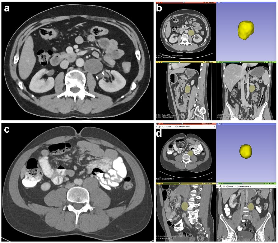
  • We assessed whether a CT radiomics–based machine learning classifier can predict histopathology of lymph nodes after post-chemotherapy lymph node dissection.
  • The trained machine learning classifier achieved a classification accuracy of 0.81 in the validation dataset with a sensitivity of 88% and a specificity of 78%, thus allowing for prediction of the presence of viable tumor or teratoma in retroperitoneal lymph node metastases.

Article: Radiomics allows for detection of benign and malignant histopathology in patients with metastatic testicular germ cell tumors prior to post-chemotherapy retroperitoneal lymph node dissection

Authors: Bettina Baessler, Tim Nestler, Daniel Pinto dos Santos, Pia Paffenholz, Vikram Zeuch, David Pfister, David Maintz & Axel Heidenreich

WRITTEN BY

  • Bettina Baeßler

    Consultant radiologist and Member of the Board of Directors of the "Machine Learning and Big Data Analysis Group", University Hospital Zurich, Department of Diagnostic and Interventional Radiology, Zurich, Switzerland

Latest posts

Become A Member Today!

You will have access to a wide range of benefits that can help you advance your career and stay up-to-date with the latest developments in the field of radiology. These benefits include access to educational resources, networking opportunities with other professionals in the field, opportunities to participate in research projects and clinical trials, and access to the latest technologies and techniques. 

Check out our different membership options.

If you don’t find a fitting membership send us an email here.

Membership

for radiologists, radiology residents, professionals of allied sciences (including radiographers/radiological technologists, nuclear medicine physicians, medical physicists, and data scientists) & professionals of allied sciences in training residing within the boundaries of Europe

  • Reduced registration fees for ECR 1
  • Reduced fees for the European School of Radiology (ESOR) 2
  • Exclusive option to participate in the European Diploma. 3
  • Free electronic access to the journal European Radiology 4
  • Content e-mails for all ESR journals
  • Updates on offers & events through our newsletters
  • Exclusive access to the ESR feed in Juisci

€ 11 /year

Yes! That is less than €1 per month.

Free membership

for radiologists, radiology residents or professionals of allied sciences engaged in practice, teaching or research residing outside Europe as well as individual qualified professionals with an interest in radiology and medical imaging who do not fulfil individual or all requirements for any other ESR membership category & former full members who have retired from all clinical practice
  • Reduced registration fees for ECR 1
  • Free electronic access to the journal European Radiology
  • Content e-mails for all 3 ESR journals 4
  • Updates on offers & events through our newsletters
  • Exclusive access to the ESR feed in Juisci

€ 0

The best things in life are free.

ESR Friends

For students, company representatives or hospital managers etc.

  • Content e-mails for all 3 ESR journals 4
  • Updates on offers & events through our newsletters

€ 0

Friendship doesn’t cost a thing.

The membership type best fitting for you will be selected automatically during the application process.

Footnotes:

01

Reduced registration fees for ECR 2024:
Provided that ESR 2023 membership is activated and approved by August 31, 2023.

Reduced registration fees for ECR 2025:
Provided that ESR 2024 membership is activated and approved by August 31, 2024.

02
Not all activities included
03
Examination based on the ESR European Training Curriculum (radiologists or radiology residents).
04
European Radiology, Insights into Imaging, European Radiology Experimental.