Radiologists with and without deep learning–based computer-aided diagnosis

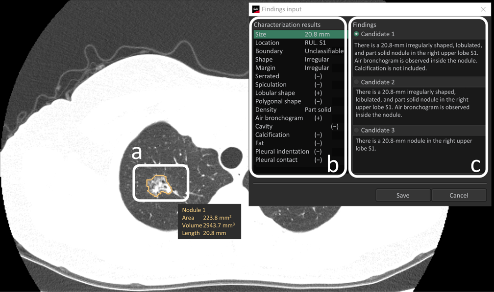
When radiologists encounter pulmonary nodules/masses in computed tomography (CT) images, they diagnose malignancy based on lesion characteristics (e.g., spiculation and calcification). However, accurate characterization requires careful observation and can be difficult, especially for inexperienced radiologists. In addition, the assessments may vary among radiologists, resulting in the low reproductivity of findings.

We investigated if commercially-available deep learning (DL)-based computer-aided diagnosis (CAD) for characterizing pulmonary nodules/masses can support radiologists. Fifteen radiologists were grouped according to their experience: L (<3 years), M (3-5 years) and H (>5 years). They observed 101 nodules/masses and evaluated the likelihoods of 15 characteristics and malignancies with and without CAD outputs. As a result, the areas under the curve (AUCs) for two items in L and M (ill-defined boundary and irregular shape in L, and ill-defined boundary and irregular margin in M), significantly improved, whereas no significant improvement was found in H. L showed the greatest increase in the AUC for diagnosis (malignancy). These results indicate CAD helps radiologists, particularly those with <5 years of experience, to characterize and diagnose pulmonary nodules/masses accurately. Moreover, intraclass correlation coefficients improved in all groups and for nearly all items, which means that CAD improved the reproductivity of findings among radiologists; the use of CAD did not prolong assessment time.

This is the first article to have proven the effectiveness of DL-based CAD for characterizing pulmonary nodules/masses. We hope for further accumulation of similar evidence in the future, clarifying a mode of implementing CAD to support radiologists efficiently.

Key points

  • Deep learning–based computer-aided diagnosis improves the accuracy of characterizing nodules/masses and diagnosing malignancy, particularly by radiologists with < 5 years of experience.
  • Computer-aided diagnosis increases not only the accuracy but also the reproducibility of the findings across radiologists.

Article: Radiologists with and without deep learning–based computer-aided diagnosis: comparison of performance and interobserver agreement for characterizing and diagnosing pulmonary nodules/masses

Authors: Tomohiro Wataya, Masahiro Yanagawa, Mitsuko Tsubamoto, Tomoharu Sato, Daiki Nishigaki, Kosuke Kita, Kazuki Yamagata, Yuki Suzuki, Akinori Hata, Shoji Kido & Noriyuki Tomiyama for the Osaka University Reading Team

WRITTEN BY

  • Shoji Kido

    Department of Artificial Intelligence Diagnostic Radiology, Osaka University Graduate School of Medicine

Latest posts

Become A Member Today!

You will have access to a wide range of benefits that can help you advance your career and stay up-to-date with the latest developments in the field of radiology. These benefits include access to educational resources, networking opportunities with other professionals in the field, opportunities to participate in research projects and clinical trials, and access to the latest technologies and techniques. 

Check out our different membership options.

If you don’t find a fitting membership send us an email here.

Membership

for radiologists, radiology residents, professionals of allied sciences (including radiographers/radiological technologists, nuclear medicine physicians, medical physicists, and data scientists) & professionals of allied sciences in training residing within the boundaries of Europe

  • Reduced registration fees for ECR 1
  • Reduced fees for the European School of Radiology (ESOR) 2
  • Exclusive option to participate in the European Diploma. 3
  • Free electronic access to the journal European Radiology 4
  • Content e-mails for all ESR journals
  • Updates on offers & events through our newsletters
  • Exclusive access to the ESR feed in Juisci

€ 11 /year

Yes! That is less than €1 per month.

Free membership

for radiologists, radiology residents or professionals of allied sciences engaged in practice, teaching or research residing outside Europe as well as individual qualified professionals with an interest in radiology and medical imaging who do not fulfil individual or all requirements for any other ESR membership category & former full members who have retired from all clinical practice
  • Reduced registration fees for ECR 1
  • Free electronic access to the journal European Radiology
  • Content e-mails for all 3 ESR journals 4
  • Updates on offers & events through our newsletters
  • Exclusive access to the ESR feed in Juisci

€ 0

The best things in life are free.

ESR Friends

For students, company representatives or hospital managers etc.

  • Content e-mails for all 3 ESR journals 4
  • Updates on offers & events through our newsletters

€ 0

Friendship doesn’t cost a thing.

The membership type best fitting for you will be selected automatically during the application process.

Footnotes:

01

Reduced registration fees for ECR 2024:
Provided that ESR 2023 membership is activated and approved by August 31, 2023.

Reduced registration fees for ECR 2025:
Provided that ESR 2024 membership is activated and approved by August 31, 2024.

02
Not all activities included
03
Examination based on the ESR European Training Curriculum (radiologists or radiology residents).
04
European Radiology, Insights into Imaging, European Radiology Experimental.