MRI radiomics features predict immuno-oncological characteristics of hepatocellular carcinoma

Advanced hepatocellular carcinoma (HCC) carries a dismal prognosis. For a decade, sorafenib, a multi-kinase inhibitor, was the only approved systemic therapy for HCC. However, its response rate in advanced HCC is only about 2%. The last few years have seen rapid approval of additional systemic therapies for HCC, including immunotherapy strategies. Immune checkpoint inhibitor nivolumab has a promising reported response rate of 20% in advanced HCC. With the emerging role of targeted therapies for advanced HCC, it also becomes increasingly relevant to accurately characterize HCC lesions in order to allow for patient-specific treatment selection. Many sophisticated techniques, including those involving immunophenotyping and genomics evaluation, have been developed. However, these methods all require invasive tissue sampling and specialized equipment and expertise.

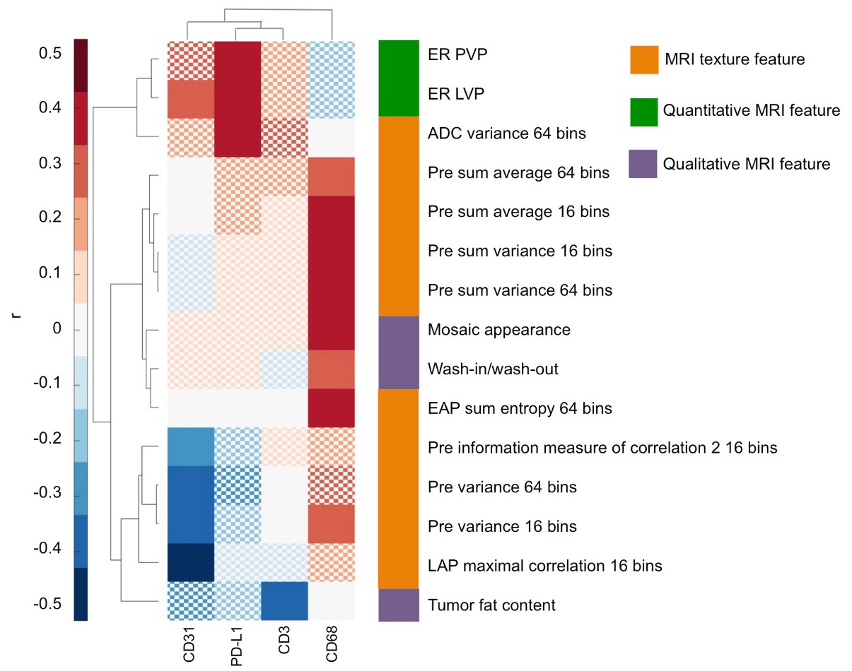
In our study, we aimed to evaluate the potential role of MRI radiomics features as surrogate noninvasive biomarkers for immuno-oncological characteristics of HCC. Radiomics is an emerging field in medical imaging, in which it is recognized that quantitative imaging features extracted from diagnostic images may be correlates of the underlying tissue biology. In our retrospective analysis of resected HCC patients with MRI performed before surgery, we found that MRI radiomics features showed significant correlation with histopathological immunophenotyping markers, including protein expression of CD3 (T-cells) and immunotherapy target PD-L1. We also found numerous significant correlations of radiomics features with RNA expression levels, including expression of immune checkpoint inhibitors PD-1 and CTLA4.

Thus, our results suggest a promising role of MRI radiomics analysis for prediction of immune-oncological features of HCC. While these results need further validation in larger and external cohorts, our study may pave a path toward personalized treatment stratification of HCC lesions using non-invasive imaging.

Key points

  • MRI radiomics features showed significant associations with immunophenotyping and genomics characteristics of hepatocellular carcinoma.
  • Radiomics features, including tumour size, showed significant associations with early hepatocellular carcinoma recurrence after resection.

Article: MRI radiomics features predict immuno-oncological characteristics of hepatocellular carcinoma

Authors: Stefanie J. Hectors, Sara Lewis, Cecilia Besa, Michael J. King, Daniela Said, Juan Putra, Stephen Ward, Takaaki Higashi, Swan Thung, Shen Yao, Ilaria Laface, Myron Schwartz, Sacha Gnjatic, Miriam Merad, Yujin Hoshida & Bachir Taouli

WRITTEN BY

  • Stefanie Hectors

    Biomedical Engineering and Imaging Institute, Department of Diagnostic, Molecular and Interventional Radiology, Icahn School of Medicine at Mount Sinai; Department of Radiology, Weill Cornell Medicine

  • Bachir Taouli

    Professor of Radiology; Director of Body MRI and Cancer Imaging Program Vice-chair of Translational Research Department of Diagnostic, Molecular and Interventional Radiology BioMedical Engineering and Imaging Institute Icahn School of Medicine at Mount Sinai

Latest posts

Become A Member Today!

You will have access to a wide range of benefits that can help you advance your career and stay up-to-date with the latest developments in the field of radiology. These benefits include access to educational resources, networking opportunities with other professionals in the field, opportunities to participate in research projects and clinical trials, and access to the latest technologies and techniques. 

Check out our different membership options.

If you don’t find a fitting membership send us an email here.

Membership

for radiologists, radiology residents, professionals of allied sciences (including radiographers/radiological technologists, nuclear medicine physicians, medical physicists, and data scientists) & professionals of allied sciences in training residing within the boundaries of Europe

  • Reduced registration fees for ECR 1
  • Reduced fees for the European School of Radiology (ESOR) 2
  • Exclusive option to participate in the European Diploma. 3
  • Free electronic access to the journal European Radiology 4
  • Content e-mails for all ESR journals
  • Updates on offers & events through our newsletters
  • Exclusive access to the ESR feed in Juisci

€ 11 /year

Yes! That is less than €1 per month.

Free membership

for radiologists, radiology residents or professionals of allied sciences engaged in practice, teaching or research residing outside Europe as well as individual qualified professionals with an interest in radiology and medical imaging who do not fulfil individual or all requirements for any other ESR membership category & former full members who have retired from all clinical practice
  • Reduced registration fees for ECR 1
  • Free electronic access to the journal European Radiology
  • Content e-mails for all 3 ESR journals 4
  • Updates on offers & events through our newsletters
  • Exclusive access to the ESR feed in Juisci

€ 0

The best things in life are free.

ESR Friends

For students, company representatives or hospital managers etc.

  • Content e-mails for all 3 ESR journals 4
  • Updates on offers & events through our newsletters

€ 0

Friendship doesn’t cost a thing.

The membership type best fitting for you will be selected automatically during the application process.

Footnotes:

01

Reduced registration fees for ECR 2024:
Provided that ESR 2023 membership is activated and approved by August 31, 2023.

Reduced registration fees for ECR 2025:
Provided that ESR 2024 membership is activated and approved by August 31, 2024.

02
Not all activities included
03
Examination based on the ESR European Training Curriculum (radiologists or radiology residents).
04
European Radiology, Insights into Imaging, European Radiology Experimental.