How machine learning-based high-dimensional CT texture analysis is influenced by segmentation margin

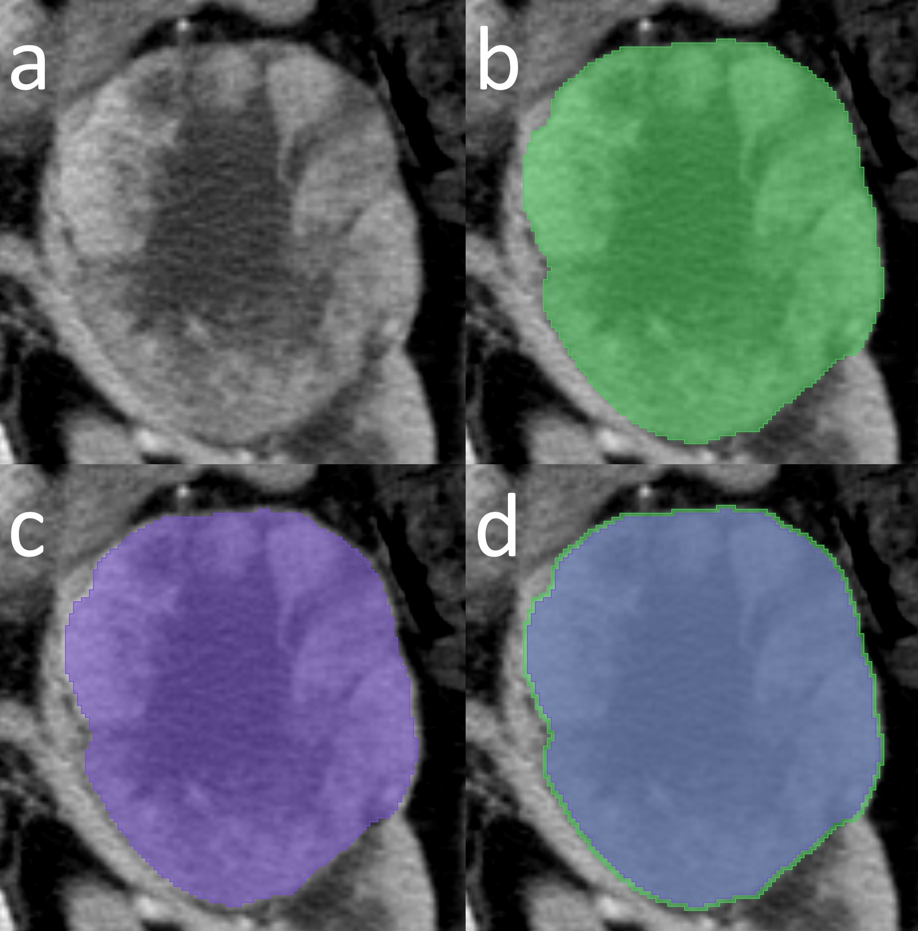
Radiomic workflows include various challenging steps. One of the most demanding steps in radiomics is the segmentation process. Particularly for the renal cell carcinomas, most of the studies used manual tumour contour delineation. In this work, our group wanted to perform an experiment by changing the segmentation margin a little bit, that is, just 2 mm, to see what happens throughout every step thereafter. We strictly preserved the nature of the data in this experiment, such as cross-validation folds, seed, and so on. We found that each step of a machine learning (ML)-based high-dimensional quantitative computed tomography texture analysis (qCT-TA) was very sensitive to even a slight change of 2 mm in segmentation margin. Despite yielding fewer texture features with excellent reproducibility, performing the segmentation focusing on the outermost boundary of the tumours provided better classification performance in ML-based qCT-TA of renal clear cell carcinomas for distinguishing nuclear grade. Although it may seem contradictory, this could be related to the quality of the information that the texture feature included in the contour-focused segmentation. In summary, findings of an ML-based high-dimensional qCT-TA may not be reproducible in clinical practice, even using the same feature selection algorithm and ML classifier, unless the possible influence of the segmentation margin is considered. We think that future research should focus on the development of highly reproducible segmentation methods for renal cell carcinomas.

Key Points

  • Each step of a machine learning (ML)-based high-dimensional quantitative computed tomography texture analysis (qCT-TA) is sensitive to even a slight change of 2 mm in segmentation margin.
  • Despite yielding fewer texture features with excellent reproducibility, performing the segmentation focusing on the outermost boundary of the tumours provides better classification performance in ML-based qCT-TA of renal clear cell carcinomas for distinguishing nuclear grade.
  • Findings of an ML-based high-dimensional qCT-TA may not be reproducible in clinical practice even using the same feature selection algorithm and ML classifier unless the possible influence of the segmentation margin is considered.

Article: Influence of segmentation margin on machine learning–based high-dimensional quantitative CT texture analysis: a reproducibility study on renal clear cell carcinomas

Authors: Burak Kocak, Ece Ates, Emine Sebnem Durmaz, Melis Baykara Ulusan, Ozgur Kilickesmez

WRITTEN BY

  • Burak Kocak

    Department of Radiology, Istanbul Training and Research Hospital

Latest posts

Become A Member Today!

You will have access to a wide range of benefits that can help you advance your career and stay up-to-date with the latest developments in the field of radiology. These benefits include access to educational resources, networking opportunities with other professionals in the field, opportunities to participate in research projects and clinical trials, and access to the latest technologies and techniques. 

Check out our different membership options.

If you don’t find a fitting membership send us an email here.

Membership

for radiologists, radiology residents, professionals of allied sciences (including radiographers/radiological technologists, nuclear medicine physicians, medical physicists, and data scientists) & professionals of allied sciences in training residing within the boundaries of Europe

  • Reduced registration fees for ECR 1
  • Reduced fees for the European School of Radiology (ESOR) 2
  • Exclusive option to participate in the European Diploma. 3
  • Free electronic access to the journal European Radiology 4
  • Content e-mails for all ESR journals
  • Updates on offers & events through our newsletters
  • Exclusive access to the ESR feed in Juisci

€ 11 /year

Yes! That is less than €1 per month.

Free membership

for radiologists, radiology residents or professionals of allied sciences engaged in practice, teaching or research residing outside Europe as well as individual qualified professionals with an interest in radiology and medical imaging who do not fulfil individual or all requirements for any other ESR membership category & former full members who have retired from all clinical practice
  • Reduced registration fees for ECR 1
  • Free electronic access to the journal European Radiology
  • Content e-mails for all 3 ESR journals 4
  • Updates on offers & events through our newsletters
  • Exclusive access to the ESR feed in Juisci

€ 0

The best things in life are free.

ESR Friends

For students, company representatives or hospital managers etc.

  • Content e-mails for all 3 ESR journals 4
  • Updates on offers & events through our newsletters

€ 0

Friendship doesn’t cost a thing.

The membership type best fitting for you will be selected automatically during the application process.

Footnotes:

01

Reduced registration fees for ECR 2024:
Provided that ESR 2023 membership is activated and approved by August 31, 2023.

Reduced registration fees for ECR 2025:
Provided that ESR 2024 membership is activated and approved by August 31, 2024.

02
Not all activities included
03
Examination based on the ESR European Training Curriculum (radiologists or radiology residents).
04
European Radiology, Insights into Imaging, European Radiology Experimental.