Automated detection of pulmonary embolism in CT pulmonary angiograms using an AI-powered algorithm

The detection of pulmonary embolism (PE) in CT pulmonary angiograms (CTPAs) is a great example of a major challenge radiologists face nowadays: The swift identification and communication of critical findings in times of sharply rising numbers of examinations performed. A potential auxiliary tool helping us to master the flood of information and separate signal (critical findings) from noise (unremarkable examinations) is algorithms that process examinations in the background and notify us in cases with suspected critical findings.

In our study, we evaluated the performance of a deep convolutional neural network (DCNN) for detection of PE using all CTPAs acquired at our institution, a university hospital, during a whole year. The algorithm based on ResNet architecture had been trained on more than 28,000 CTPAs from other institutions. Among others, we found a sensitivity of 92.7% and a specificity of 95.5% on a per-examination level, which makes it a valuable aide for radiologists.

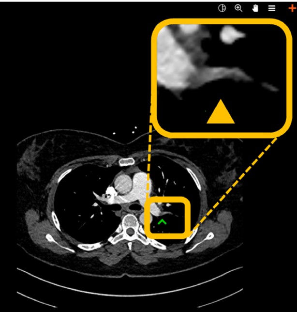
While these kinds of performance checks on real clinical data are indispensable, they are just one of many steps to meaningful application of AI in radiology. Starting now, we should widen our focus and include two more aspects: First, how exactly do we integrate algorithm results in clinical workflows so they make lives of radiologists easier and don’t add extra workload. This is not only key for acceptance of AI, but also its practical implementability. At our department, we opted for a widget-based solution for the PE detection algorithm: A pop-up window occurs at the radiologist’s workstation whenever PE is suspected (and only then). It includes a key image of the suspected embolus- prompting direct reading of the case by a radiologist. And second, we need to take the impact on patient outcomes into account: the best performing algorithm is worthless if its results do not find their way to the right person at the right time.

Staying with our example, this means we need – among other things – to have a closer look at the influence of the algorithm on communication times to referring physicians. The work has just begun.

Key points

  • An AI-based prototype algorithm showed a high degree of diagnostic accuracy for the detection of pulmonary embolism on CTPAs.
  • It can therefore help clinicians to automatically prioritize exams with a high suspection of pulmonary embolism and serve as secondary reading tool.
  • By complementing traditional ways of worklist prioritization in radiology departments, this can speed up the diagnostic and therapeutic workup of patients with pulmonary embolism and help to avoid false negative calls.

Article: Automated detection of pulmonary embolism in CT pulmonary angiograms using an AI-powered algorithm

Authors: Thomas Weikert, David J. Winkel, Jens Bremerich, Bram Stieltjes, Victor Parmar, Alexander W. Sauter & Gregor Sommer

WRITTEN BY

  • Thomas Weikert

    Clinic of Radiology & Nuclear Medicine, University Hospital Basel, Switzerland

Latest posts

Become A Member Today!

You will have access to a wide range of benefits that can help you advance your career and stay up-to-date with the latest developments in the field of radiology. These benefits include access to educational resources, networking opportunities with other professionals in the field, opportunities to participate in research projects and clinical trials, and access to the latest technologies and techniques. 

Check out our different membership options.

If you don’t find a fitting membership send us an email here.

Membership

for radiologists, radiology residents, professionals of allied sciences (including radiographers/radiological technologists, nuclear medicine physicians, medical physicists, and data scientists) & professionals of allied sciences in training residing within the boundaries of Europe

  • Reduced registration fees for ECR 1
  • Reduced fees for the European School of Radiology (ESOR) 2
  • Exclusive option to participate in the European Diploma. 3
  • Free electronic access to the journal European Radiology 4
  • Content e-mails for all ESR journals
  • Updates on offers & events through our newsletters
  • Exclusive access to the ESR feed in Juisci

€ 11 /year

Yes! That is less than €1 per month.

Free membership

for radiologists, radiology residents or professionals of allied sciences engaged in practice, teaching or research residing outside Europe as well as individual qualified professionals with an interest in radiology and medical imaging who do not fulfil individual or all requirements for any other ESR membership category & former full members who have retired from all clinical practice
  • Reduced registration fees for ECR 1
  • Free electronic access to the journal European Radiology
  • Content e-mails for all 3 ESR journals 4
  • Updates on offers & events through our newsletters
  • Exclusive access to the ESR feed in Juisci

€ 0

The best things in life are free.

ESR Friends

For students, company representatives or hospital managers etc.

  • Content e-mails for all 3 ESR journals 4
  • Updates on offers & events through our newsletters

€ 0

Friendship doesn’t cost a thing.

The membership type best fitting for you will be selected automatically during the application process.

Footnotes:

01

Reduced registration fees for ECR 2024:
Provided that ESR 2023 membership is activated and approved by August 31, 2023.

Reduced registration fees for ECR 2025:
Provided that ESR 2024 membership is activated and approved by August 31, 2024.

02
Not all activities included
03
Examination based on the ESR European Training Curriculum (radiologists or radiology residents).
04
European Radiology, Insights into Imaging, European Radiology Experimental.