A fully automated software platform for structural mitral valve analysis

There has been great interest in the development and application of artificial intelligence (AI) in the field of medical imaging. AI has the potential to be useful in multiple arenas including the improvement of diagnostic accuracy as well as improving workflow and helping increase human efficiency in study interpretation. Cardiac imaging has been one focus of AI and has yielded promising results, such as aortic valve analysis in the setting of TAVR evaluation and non-invasive CT derived fractional flow reserve in the assessment of obstructive coronary artery disease, to name a few. However, to date, this has not been the case for the mitral valve, largely owing to its complex anatomy. True 3-D measurement of the mitral annulus (diameter, area, circumference) utilizing CT imaging can be difficult and time-consuming. At the same time, we are seeing advancement and increasing options for percutaneous approaches to mitral valve interventions, making accurate, non-invasive assessment of mitral valve anatomy a necessity. Thus, a fully automated and accurate analysis tool for the mitral valve would be an attractive innovation for deployment in the clinical arena.

We compared a fully automated mitral valve assessment prototype to a hybrid user-driven software package in the assessment of several mitral valve sizing parameters. Additionally, we determined the time needed to obtain these measurements between the two techniques. The fully automated software demonstrated a significant reduction in time to results; however, correlation was suboptimal. When adding manual adjustments to the fully automated prototype, correlation improved, yielding good to very good correlation when compared to the user-driven analysis.

While showing great results on our patient cohort under study conditions, the automated software struggled in the setting of severe valve calcification and motion artefact. The fully automated software demonstrated systematic under-sizing compared to the user-driven package. Manual adjustments were needed to improve correlation, which resulted in less robust time savings. While a fully automated mitral valve assessment package is an attractive prospect, current iterations are not ready for integration into clinical practice and will require further development and refinement.

Key points

  • The novel software platform allows for a fully automated analysis of mitral valve structures.
  • An excellent agreement was found between the fully automated measurement with manual adjustments and the user-driven analysis.
  • The software showed quicker measurement time compared with the standard analysis of the mitral valve.

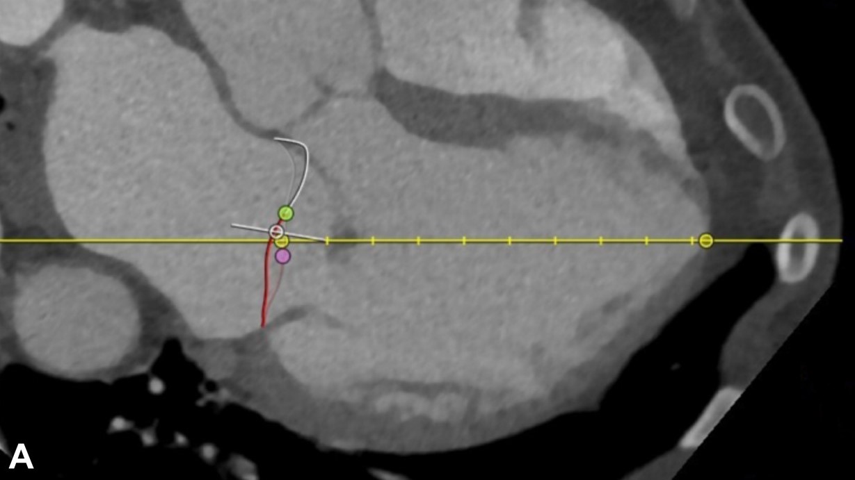
Article: A fully automated software platform for structural mitral valve analysis

Authors: Robert Steinbach, U. Joseph Schoepf, L. Parkwood Griffith, Marly van Assen, Matthias Renker, Pooyan Sahbaee, Chris Schwemmer, Andreas M. Fischer, Akos Varga-Szemes, Simon S. Martin & Richard R. Bayer II

WRITTEN BY

  • U. Joseph Schoepf

    Division of Cardiovascular Imaging, Department of Radiology and Radiological Science, Medical University of South Carolina, Charleston, South Carolina, USA

Latest posts

Become A Member Today!

You will have access to a wide range of benefits that can help you advance your career and stay up-to-date with the latest developments in the field of radiology. These benefits include access to educational resources, networking opportunities with other professionals in the field, opportunities to participate in research projects and clinical trials, and access to the latest technologies and techniques. 

Check out our different membership options.

If you don’t find a fitting membership send us an email here.

Membership

for radiologists, radiology residents, professionals of allied sciences (including radiographers/radiological technologists, nuclear medicine physicians, medical physicists, and data scientists) & professionals of allied sciences in training residing within the boundaries of Europe

  • Reduced registration fees for ECR 1
  • Reduced fees for the European School of Radiology (ESOR) 2
  • Exclusive option to participate in the European Diploma. 3
  • Free electronic access to the journal European Radiology 4
  • Content e-mails for all ESR journals
  • Updates on offers & events through our newsletters
  • Exclusive access to the ESR feed in Juisci

€ 11 /year

Yes! That is less than €1 per month.

Free membership

for radiologists, radiology residents or professionals of allied sciences engaged in practice, teaching or research residing outside Europe as well as individual qualified professionals with an interest in radiology and medical imaging who do not fulfil individual or all requirements for any other ESR membership category & former full members who have retired from all clinical practice
  • Reduced registration fees for ECR 1
  • Free electronic access to the journal European Radiology
  • Content e-mails for all 3 ESR journals 4
  • Updates on offers & events through our newsletters
  • Exclusive access to the ESR feed in Juisci

€ 0

The best things in life are free.

ESR Friends

For students, company representatives or hospital managers etc.

  • Content e-mails for all 3 ESR journals 4
  • Updates on offers & events through our newsletters

€ 0

Friendship doesn’t cost a thing.

The membership type best fitting for you will be selected automatically during the application process.

Footnotes:

01

Reduced registration fees for ECR 2024:
Provided that ESR 2023 membership is activated and approved by August 31, 2023.

Reduced registration fees for ECR 2025:
Provided that ESR 2024 membership is activated and approved by August 31, 2024.

02
Not all activities included
03
Examination based on the ESR European Training Curriculum (radiologists or radiology residents).
04
European Radiology, Insights into Imaging, European Radiology Experimental.